Visual diagnosis of symptoms and laboratory confirmation
Content generated by artificial intelligence — verify before application
This article was automatically compiled from a collection of hazelnut literature using artificial intelligence and may contain errors or outdated data. It serves as a starting point for further research, not as professional advice. Dosages, products, and timing are informative and depend on the cultivar, location, and current regulations — always consult an advisory service and official sources before application.
Proper protection of hazelnut (Corylus avellana L.) orchards begins with correct diagnosis. Before a producer reaches for any treatment, they must know what they are actually looking at: is the browning of a bud the result of a fungal infection, bacterial canker, frost, or simply a physiological disorder in nutrition? An incorrect diagnosis leads to a double loss — wasted cost of spraying and missed timing for an effective measure. This article provides a systematic approach to "reading" symptoms on buds, leaves, shoots, and fruit, explains how to distinguish between biotic (living pathogens) and abiotic (non-living factors) causes, and describes how and when to seek laboratory confirmation of the pathogen.
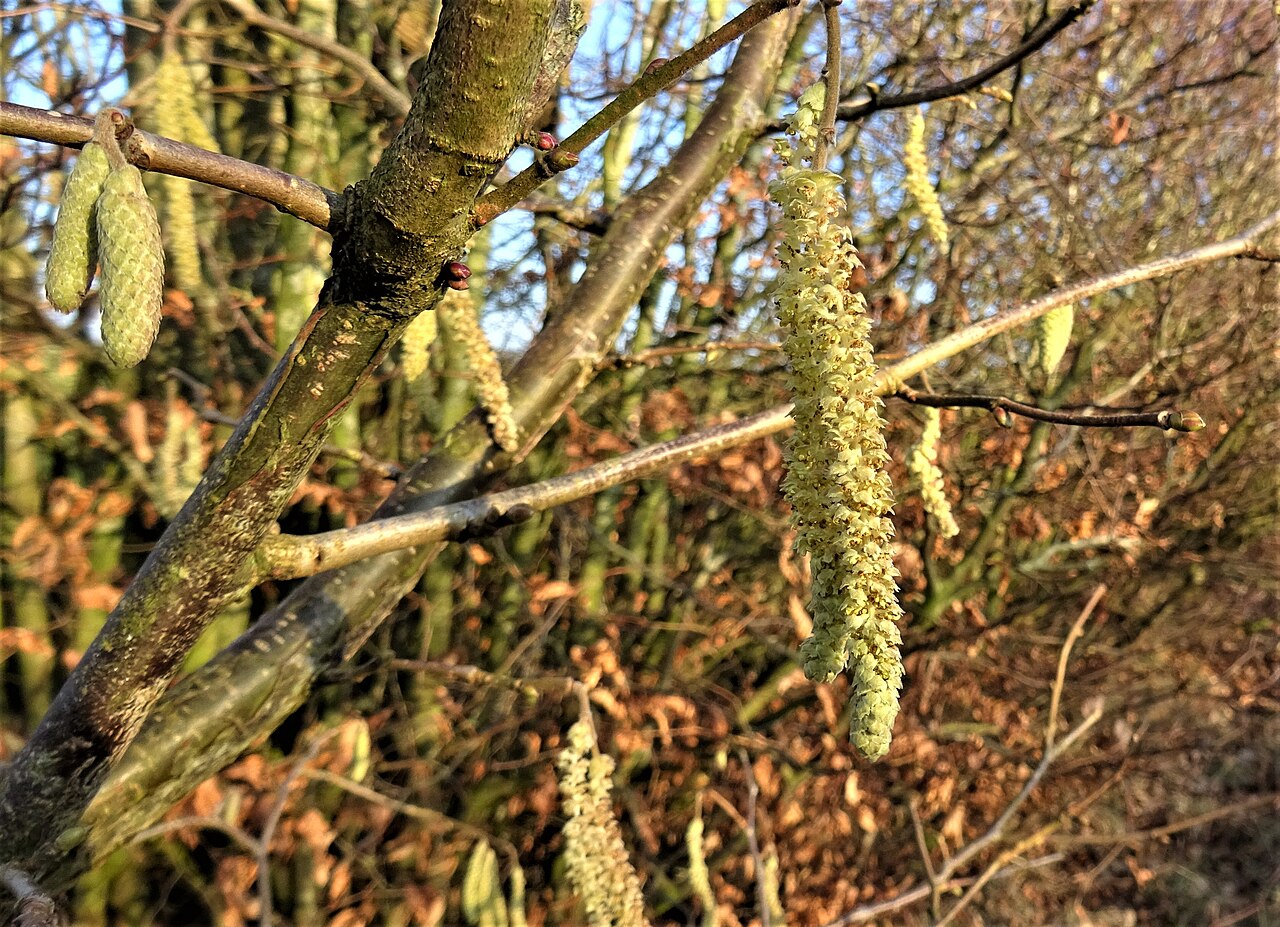
Diagnostic principle: symptom is not cause
The biggest trap in field diagnosis is equating the symptom (visible changes on the plant) with the cause. The same symptom — for example, browning and drying of buds — can appear due to several different causes: the fungus Gloeosporium coryli (syn. Monostichella coryli), the bacterium Pseudomonas syringae pv. avellanae, winter frost damage, or water deficiency. The task of the diagnostician is not to stop at describing the change, but to go one step further and ask: which set of signs explains this?
In phytopathology, two terms are distinguished:
- Symptoms — the reaction of the plant itself (yellowing, necrosis, wilting, stunting, shedding).
- Signs — visible parts of the pathogen itself (mycelium, fungal fruiting bodies, bacterial exudate, eggs, or adult pests).
Signs are more diagnostically valuable than symptoms because they directly point to the organism. Black dots (acervuli) on leaf spots, powdery mildew coating, or droplets of red exudate on branches are strong clues that narrow down the list of suspects.
Five questions before every diagnosis
- Distribution in the orchard — is the problem random (often abiotic, e.g., frost, soil) or spreading in patches from tree to tree (often biotic, infectious)?
- Temporal sequence — when did it appear and does it follow a weather event (heavy rain, frost, drought, hail)?
- Which organs are affected — only fruit, only leaves, or everything at once?
- Are there signs of the pathogen — fruiting bodies, coating, exudate, pest?
- Pattern within the plant — does it start from the shoot tip downwards, from the leaf edge towards the center, or from the root/trunk?
Systematic reading of symptoms by organ
A disciplined inspection goes in order: bud → leaf → shoot/branch → fruit → root/trunk. This ensures nothing is missed and it is easier to piece together a complete picture.
Bud
The bud is often the first indicator of trouble because both infectious diseases and frost manifest on it.
- Browning and drying of sprouting buds and young shoots — typical for the fungus Gloeosporium / Monostichella coryli (leaf spot and bud drying). The fungus often spreads into the shoot, which dries out; stunted shoots can develop from infected buds. Symptoms intensify after prolonged cold and rainy periods during the growing season.
- Brown, soft buds that do not leaf out — a classic early sign of bacterial canker / blight of hazelnut (Pseudomonas syringae pv. avellanae; related to Xanthomonas arboricola pv. corylina). A very noticeable accompanying sign is stunted male flowers (catkins).
- Deformed, swollen ("gourd-shaped") buds — indicate hazelnut bud gall mite (Phytoptus avellanae), not a disease; this is a sign of a pest.
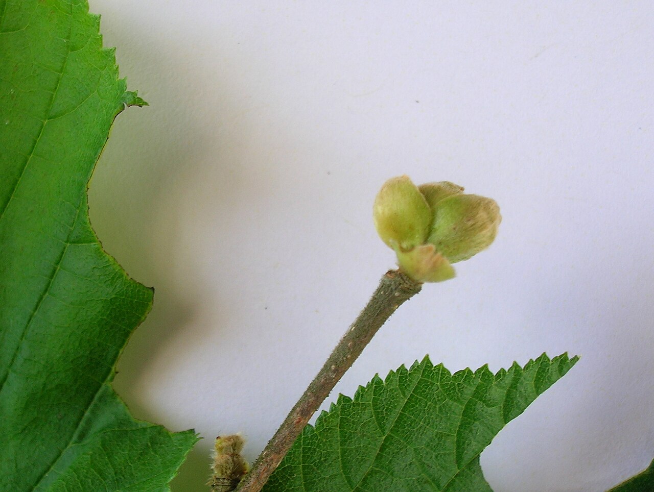
- A bud that does not sprout after a mild winter, without signs of a pathogen — consider frost or a physiological cause before fungus/bacteria.
Leaf
The leaf is a large "board" on which much is written.
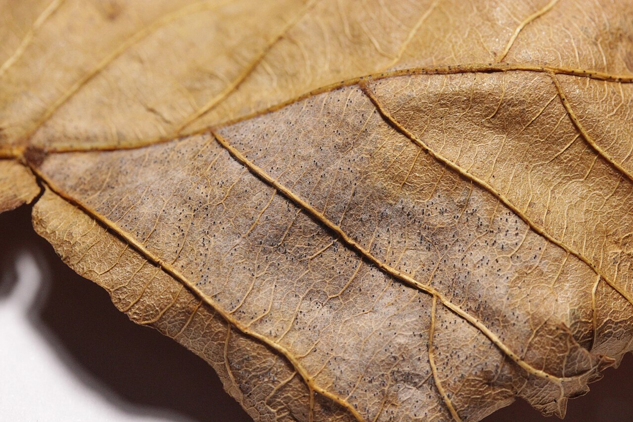
- Round spots up to 2 cm with a lightened ring-like zone bordered by a darker brown zone, late summer and early autumn — leaf spot (Gloeosporium / Monostichella coryli). Spots merge, affecting the tissue between veins, necrosis occurs, and the leaf falls off. Black dots (acervuli) — signs of the fungus — are visible to the naked eye on the spots.
- Powdery coating, initially on the underside of the leaf (lower side), and powdery spots on the upper side — hazelnut powdery mildew (Phyllactinia guttata, and more recently Erysiphe corylacearum). Leaves yellow and may shed (defoliation). Symptoms typically appear from August to October, and earlier in humid years (relative humidity 70–80%).
- "Wet", shiny leaves due to honeydew — a sign of a strong population of aphids (Myzocallis spp. and others); honeydew drips from the underside of upper leaves onto the upper side of lower leaves, and sooty mold later develops on it.
- Light green color and loss of turgor, visible from late July, accompanied by delayed vegetation and lack of growth — possible wilting (verticillium), Verticillium spp.
- Mosaic patterns, lightening along veins — possible viruses (e.g., apple mosaic virus, ApMV), which can only be reliably confirmed in the laboratory.
- Sunscald on leaf edges/plates and on bracts of the fruit — abiotic cause (heat/light stress), without signs of a pathogen.
Shoot and branch
- Drying of shoots continuing from an infected bud — fungal leaf spot/bud drying has spread into the shoot.
- Dark depressions on the bark that crack, bark falls off down to the wood, first symptoms in spring — bark canker (Neonectria ditissima). If a large area is affected, the part of the branch above the zone dries out.
- Breaking of skeletal (branching) branches with droplets of red exudate at the infected site — a typical symptom of fungal canker (Cytospora decipiens), recorded in Croatian orchards.
- Root suckers that quickly decay, spread of infection from branches and trunk to the root — pattern of spread in bacterial canker (P. syringae pv. avellanae).
Fruit
The fruit is the most economically important organ, and simultaneously the place where pests and diseases overlap.
-
Yellowing of the fruit from the tip, then browning of the shell (woody outer layer) and changes in the kernel — kernel rot caused by the fungus Nematospora coryli. Key diagnostic detail: if the infection is the result of a fungus transmitted by a bug sting, brown droplets are visible on the inside of the shell; if the absence of the seed is the result of a non-parasitic (abiotic) factor or poor pollination, there is no droplet.
-
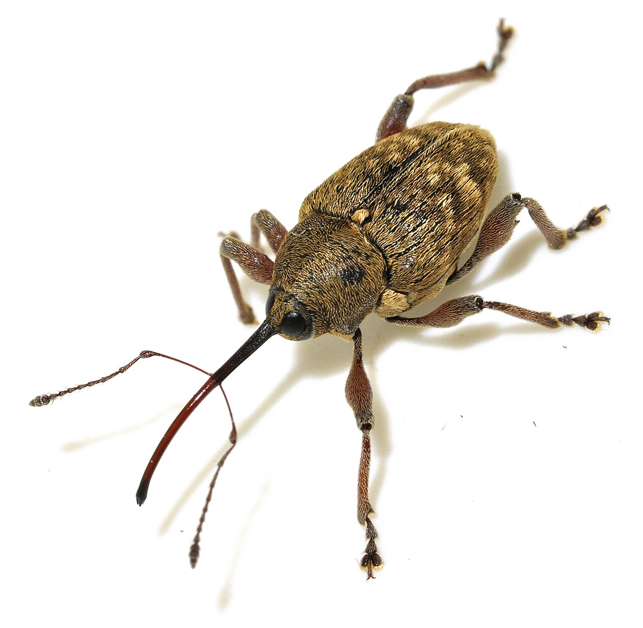
Whitish spots at the sting site, hollow shell, "traumatic abortion" — early attack by bugs (May–June, while the kernel is just forming); the kernel turns black and dries out. Later attack results in yellow to brown, sour kernels with an unpleasant smell, unsuitable for eating and processing.
- Light brown to chestnut spots on the pericarp that spread over the entire surface of the fruit; early-infected fruits fall off — brown rot of fruit (Monilinia laxa, Monilinia spp.).
- Brown spots on the shell and browning of the kernel after heavy rains in May/June — a combination of gray spot, fruit rot, and damage from bugs/hazelnut weevil; often poor pollination as a concurrent abiotic cause.
Root and trunk
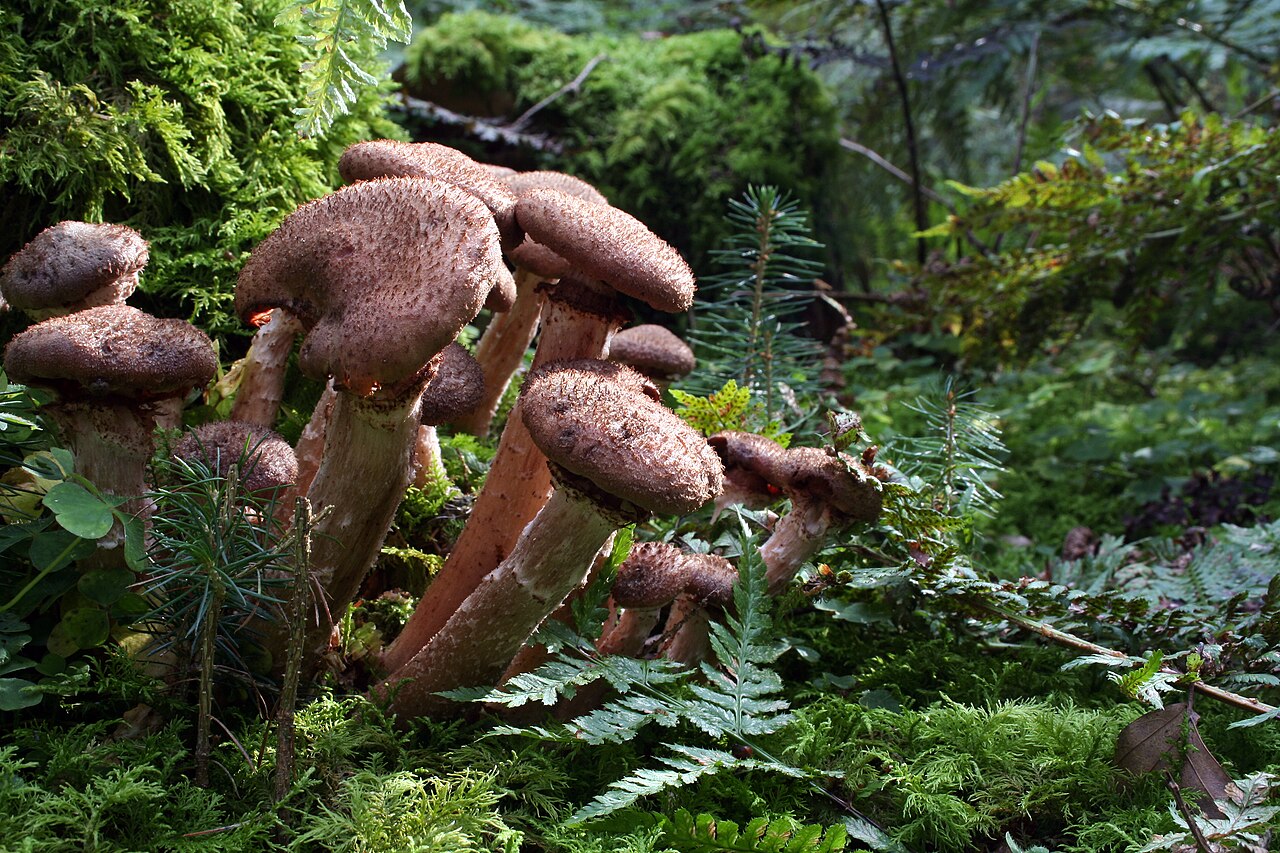
- Drying of the entire shrub or tree, root rot — Armillaria mellea (honey fungus); infection through flowers, lenticels, and wounds in spring and autumn.
- Gradual decay with wilting leaves during summer — verticillium wilt (Verticillium spp.).
Distinguishing biotic and abiotic causes
This is the central skill of diagnosis. A mistake at this step means spraying against a "disease" that doesn't exist, or ignoring an infection that is spreading.
| Feature | Biotic cause (living: fungus, bacterium, virus, pest) | Abiotic cause (non-living: frost, drought, heat, nutrition, soil) |
|---|---|---|
| Distribution in the orchard | Often in patches, spreads from tree to tree, from edges inward | Often uniform or related to terrain configuration (frost pockets, dry/compacted soil) |
| Temporal course | Progresses during the season, "jumps" to neighboring plants | Sudden appearance linked to an event (frost, hail, heatwave) |
| Signs of the pathogen | Present (mycelium, fruiting bodies, exudate, pest, eggs) | Absent |
| Pattern on the plant | Often uneven, on specific organs/sides | Symmetrical (e.g., both sides equally, all shoot tips) |
| Transition healthy–sick | Often sharp boundary, sometimes a halo | Gradual transition |
| Reaction to neighboring species | Specific host (hazelnut) | Affects other species in the vicinity (e.g., frost, hail) |
Practical examples of overlap in hazelnut:
- Browned buds: biotic (fungus, bacterium) if they spread and are accompanied by shoot drying/stunted catkins; abiotic (frost) if they appeared suddenly after cold, uniformly, and without signs of a pathogen.
- Empty or unfilled kernel: bug (biotic) leaves brown droplets in the shell; poor pollination or stress (abiotic) leaves no droplets.
- Leaf drying at the edge: heat/salt stress or drought (abiotic) is usually symmetrical; infection (biotic) often has spots and signs of fungus.
Golden rule
When there are no signs of a pathogen, and the pattern is symmetrical and linked to a weather event — first suspect an abiotic cause. Do not spray fungicide against frost.
When and how to send a sample to the laboratory
Field diagnosis has its limits. Laboratory confirmation is necessary when:
- symptoms are ambiguous or overlapping (e.g., bacterial canker vs. fungal canker vs. frost damage),
- there is suspicion of a quarantine or regulated organism (e.g., Xanthomonas arboricola pv. corylina),
- the disease is spreading rapidly and the cause is unclear,
- there is suspicion of a virus (cannot be reliably confirmed visually),
- certification of planting material is planned or new hazelnut trees are being introduced.
In Croatia, for testing leaves, twigs, and nuts for diseases, you can contact the competent plant protection institute and the advisory service; nutrient analyses from leaves and soil are performed by specialized (and private) laboratories. Always check the current contacts and sample submission protocols with the competent institution before sending samples.
How to collect and prepare a sample (field steps)
The principles apply generally to plant diagnostics and are also covered by guidelines from public diagnostic clinics:
- Collect a representative sample that includes both diseased and transitional (border) tissue — a mixture of symptomatic and apparently healthy parts. Dead, completely decomposed tissue is worthless because it is overgrown by secondary microflora.
- For branches/shoots, take a piece at the transition between necrotic and healthy tissue.
- Collect the sample fresh; do not dry it or place it in water.
- Wrap in a dry paper towel (absorbs moisture and slows decay), place in a zip-lock plastic bag, and preferably in a second bag.
- Keep in the refrigerator (not the freezer) until shipping; send as soon as possible, preferably at the beginning of the week and via express delivery if possible.
- Attach a completed form with data: cultivar, location (GPS/plot), orchard age, date of appearance, symptom distribution, affected organs, weather conditions, treatment history, and suspicion.
| Organ / Target | What to collect | Note |
|---|---|---|
| Shoot dieback, canker | 10–15 cm twigs with healthy–diseased transition | Do not collect completely dry wood |
| Leaf spot / powdery mildew | 10+ leaves with various stages of spotting | Include both fresh and older spots |
| Fruit/nut rot | Several nuts with varying degrees of damage | Preferably also apparently healthy ones for comparison |
| Virus suspicion | Young leaves with symptoms | Keep cooled, deliver quickly |
| Nutrient analysis | Leaf sample per protocol (number, position, timing) | Follow laboratory instructions exactly |
Laboratory confirmation methods
Understanding the methods helps in interpreting results:
| Method | What it does | Typical application in hazelnut |
|---|---|---|
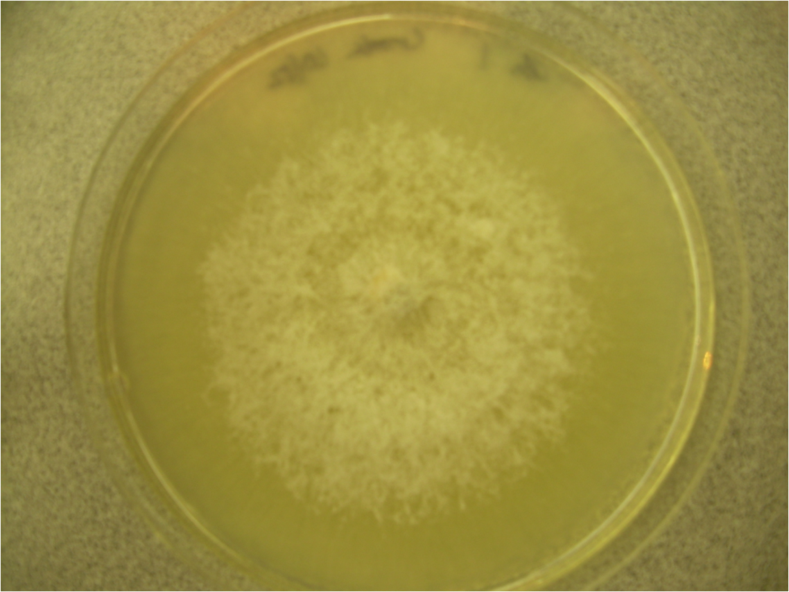
| Isolation on culture medium (e.g., KDA – potato dextrose agar) | Cultivation of the pathogen from tissue; identification by colony appearance and microscopy | Fungal pathogens (e.g., from twigs with necrosis) |
| Surface disinfection before isolation | Removes secondary flora (e.g., ~5 % NaOCl for a few minutes, rinsing with sterile water) | Preparation of tissue fragments for isolation |
| Pathogenicity test (inoculation) | Proves that the isolate actually causes the disease (Koch's postulates) | Confirmation of the isolate's role |
| ELISA (enzyme-linked immunosorbent assay) | Detects antigens/proteins of the pathogen | Viruses (e.g., ApMV); historically also for the fungus A. anomala |
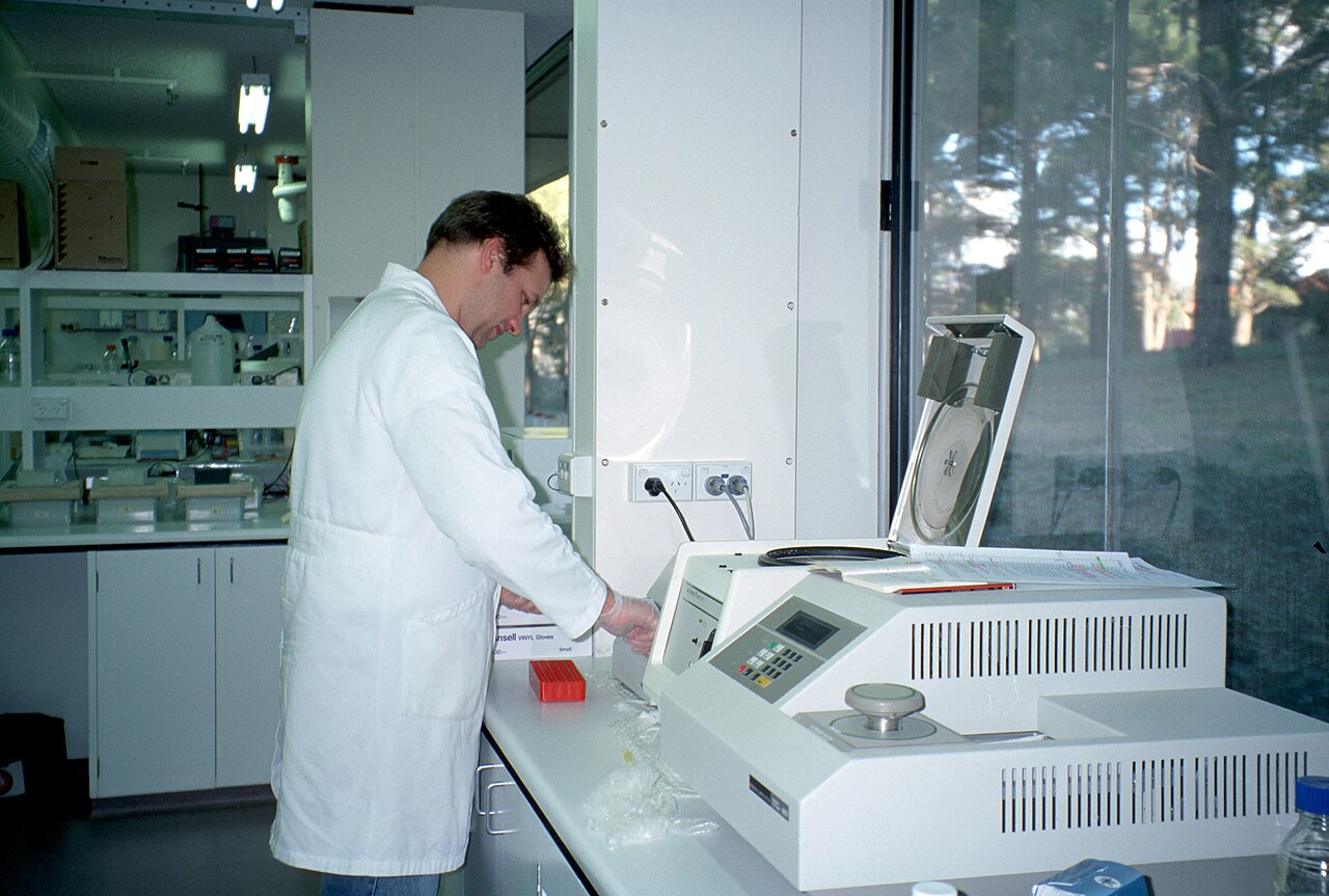
| PCR / Real-time PCR, sequencing | Molecular detection of pathogen DNA/RNA; highly specific | Confirmation of bacteria (Xac), viruses, fungal species |
| LAMP (isothermal amplification) | Rapid molecular method, applicable closer to the field | Developed for detecting powdery mildew Erysiphe corylacearum |
The values and concentrations mentioned above (e.g., NaOCl content, incubation temperature) are informative and taken from literature; the specific protocol is determined by the laboratory. Typically, for molecular confirmation, the percentage of nucleotide identity with a reference strain is also reported (e.g., Croatian isolates of Xanthomonas arboricola pv. corylina showed 99.88–100 % identity with a Spanish strain), illustrating the precision of molecular identification.
Practical recommendations and common mistakes
- Monitor the orchard regularly — monthly inspections with notes and photos (as in documented Croatian research) allow you to catch the early stage and spread pattern.
- Photograph the symptom in context — whole plant, then organ, then detail of the sign; include date and location.
- Do not diagnose based on a single leaf — one symptom is rarely sufficient; look for a set of signs and distribution.
- Do not spray "just in case" — without a diagnosis, you waste money and can worsen resistance.
- Distinguish cause from effect — bacterioses are often causally linked to previous damage from insects (bugs, hazelnut weevil) that create entry points; controlling insects is often the first step against fruit rot.
- Suspected quarantine organism = notify the competent authority — do not move planting material.
- Maintain tool hygiene — disinfecting pruners between trees prevents the spread of canker and bacterioses.
- Send laboratory samples fresh and cooled — the most common mistake is sending dried or completely rotten samples from which the true pathogen cannot be isolated.
Gallery
Sources
This article is an AI summary compiled from an internal literature collection on hazelnut (RAG corpus) and supplemented with public sources. Main sources used:
- D. Bošnjak — Hazelnut (manual on diseases and pests, agro-klaster.hr) — https://agro-klaster.hr/wp-content/uploads/2024/01/dr.sc_.-D.-Bo%C5%A1njak-Lijeska-2.pdf
- Diseases and pests of hazelnut (final/thesis works, Repozitorij) — leaf spot and bud dieback, brown fruit rot, kernel rot, powdery mildew, bugs
- Plavec et al. (2024) — Xanthomonas arboricola pv. corylina, causal agent of bacterial blight of hazelnut in nurseries and orchards in Croatia (molecular detection, PCR)
- Morphological and pathogenic characterization — isolation of pathogen from hazelnut twigs (KDA, NaOCl, incubation) — https://doi.org/10.52450/zrmg05
- Occurrence and diversity of apple mosaic ilarvirus in apple and hazelnut in Serbia — ELISA and RT-PCR detection of ApMV
- Notifications and recommendations for hazelnut orchard owners (advisory service) — gray spots, brown fruit rots, registered products (Signum, Champion WG, Calypso SC, etc.)
- Hazelnut Cultivation (Poljoprivredni Forum) — availability of diagnostics (plant protection institute, advisory service, private laboratories) and sample collection
- Development of a Field-Deployable LAMP Assay for Erysiphe corylacearum in Hazelnut — https://www.ncbi.nlm.nih.gov/pmc/articles/PMC12842720/
- Pseudomonas syringae-Associated Hazelnut Decline in Central Italy — https://www.ncbi.nlm.nih.gov/pmc/articles/PMC4739619/
- Plant Molecular Diagnostic Lab (UGA) — instructions for sample collection and submission — https://site.caes.uga.edu/mdl/instructions-for-sample-collection-and-submisson/
- Texas Plant Disease Diagnostic Lab — plant sampling — https://plantclinic.tamu.edu/plant-sampling/
- CSU Extension — Molecular Methods for Diagnosing Plant Diseases (ELISA, PCR, RPA, LAMP, NGS) — https://extension.colostate.edu/topic-areas/agriculture/molecular-methods-for-diagnosing-plant-diseases/